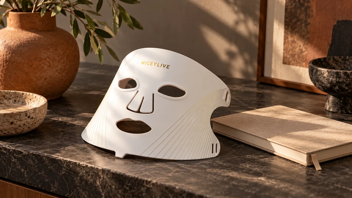
The Cellular Science: How Red and NIR Photons Trigger Neocollagenesis
At MICEYLIVE, we help you understand face and body light-care formats before choosing a device.
Light-emitting diode (LED) facial therapy operates on the established biomedical mechanism of photobiomodulation (PBM). Unlike invasive cosmetic procedures or ablative laser resurfacing technologies that force skin regeneration through thermal coagulative injury, photobiomodulation is an entirely non-thermal and non-ablative photochemical process. When calibrated wavelengths of visible red and invisible near-infrared light irradiate human facial tissue, optical photons penetrate through epidermal layers to reach dermal fibroblasts. Within these fibroblasts, photons are selectively absorbed by unit IV of the mitochondrial respiratory electron transport chain: the primary photoacceptor enzyme known as cytochrome c oxidase. This photonic absorption accelerates electron transport, elevates mitochondrial membrane potential, and drives an immediate surge in intracellular adenosine triphosphate (ATP) synthesis.
The metabolic influx of ATP, combined with the transient modulation of reactive oxygen species and intracellular signaling messengers, stimulates dermal fibroblasts at the transcriptional level. Cellular reverse transcription-polymerase chain reaction (RT-PCR) analyses confirm that photobiomodulation triggers marked upregulation of Type I procollagen mRNA expression. Dermal fibroblasts are stimulated to synthesize and secrete soluble procollagen precursor polypeptides, initiating the complex biological cascade required to replenish the extracellular matrix. Modern home-use LED masks deliver these therapeutic photon fluences through narrow-band semiconductor arrays, safely avoiding harmful ultraviolet radiation and thermal damage.
Key Takeaways: Realistic LED Wrinkle Reduction at a Glance
-
Non-Thermal Cellular Mechanism: Red (630-660 nm) and near-infrared (830-850 nm) light stimulate mitochondrial cytochrome c oxidase and ATP synthesis to upregulate Type I procollagen production without thermal damage.
-
Proven Ultrasound Collagen Density Increase: Controlled clinical trials using 22 MHz ultrasonography demonstrate a statistically significant increase in reticular dermal collagen density (p < 0.001) following 30 sessions over 10 to 12 weeks.
-
Quantifiable Skin Roughness Reduction: Objective 3D optical profilometry confirms a 20% to 35% reduction in skin roughness scores and wrinkle volume across lateral periorbital and facial areas.
-
The 12-Week Biological Timeline: Meaningful visible softening of fine lines requires 8 to 12 weeks of disciplined adherence; microscopic collagen synthesis takes 60 to 90 days to reorganize into dense extracellular bundles.
-
Audit Marketing Photography: Disregard dramatic 14-day social media transformations; deceptive before-and-after photos exploit directional overhead down-lighting shadows, wide-angle lens distortion, and topical humectants.
-
Consistency and Maintenance: Maximum therapeutic benefit requires 3 to 5 sessions per week (10 to 15 minutes per session), followed by ongoing weekly maintenance to prevent collagen regression.
Dual-Wavelength Synergy: Papillary vs. Reticular Dermal Penetration
The clinical efficacy of an LED face mask for wrinkle reduction is physically governed by tissue penetration optics. Human skin is a turbid optical medium characterized by high optical scattering and absorption from endogenous chromophores, particularly epidermal melanin and capillary hemoglobin. Short-wavelength visible light suffers extreme Rayleigh scattering, severely limiting penetration into the skin. Visible red light, calibrated between 630 nm and 660 nm (peaked clinically at 633 nm), exhibits lower optical scattering, allowing photons to penetrate 1 to 2 millimeters through the dermal-epidermal junction into the papillary and upper reticular dermis. In this zone, 633 nm red light directly stimulates sub-epidermal fibroblasts, addressing micro-roughness, superficial crepiness, and early fine lines.
To achieve structural wrinkle smoothing, however, photon energy must penetrate deeper into the thick reticular dermis. Near-infrared (NIR) light, centered precisely at 830 nm (ranging from 830 nm to 850 nm), lies within the optical therapeutic window where tissue scattering and melanin absorption reach their lowest points. Photons at 830 nm penetrate 5 to 10 millimeters into deep reticular connective tissue, subcutaneous structures, and superficial vascular networks. While near-infrared light is invisible to the human eye, its biophysical actions stimulate deep perivascular fibroblasts, optimize capillary perfusion, and accelerate extracellular matrix reorganization. When 633 nm red light and 830 nm near-infrared light are administered simultaneously in dual-wavelength arrays, the skin receives synergistic multi-depth photostimulation that superficial red light alone cannot achieve.
Objective Clinical Trial Metrics: Ultrasonography and Profilometry Data
Evaluating before-and-after wrinkle reduction requires examining validated medical trial metrics rather than subjective aesthetic impressions. In a landmark randomized controlled clinical trial conducted by Wunsch and Matuschka (2014), 113 subjects were enrolled to investigate the anti-aging efficacy of red and near-infrared light phototherapy. Over a 10 to 12 week period, subjects received 30 photobiomodulation sessions administered twice weekly. To objectively evaluate structural changes beneath the skin surface, investigators employed high-frequency 22 MHz ultrasonography, which quantifies acoustic reflection density in the reticular dermis. The ultrasound scans revealed a statistically significant increase in intradermal collagen density (p < 0.001) in treated subjects compared to untreated control groups, providing direct physical evidence of light-induced neocollagenesis.
In parallel with ultrasound acoustic measurements, clinical researchers utilize computerized 3D optical profilometry to evaluate surface micro-topography. Optical profilometry scans skin surface roughness, measuring microscopic peaks and valleys across facial wrinkles before and after treatment courses. In the Wunsch and Matuschka trial, profilometric evaluations recorded a statistically significant reduction in facial skin roughness ranging between 20% and 35% across peri-orbital and lateral temporal test areas. Similarly, a comprehensive clinical evaluation by Russell et al. (2005) assessing combination 633 nm red and 830 nm near-infrared LED therapy demonstrated marked reductions in wrinkle depth and fine line severity. Independent dermatologist evaluations in the Russell study recorded 52% of subjects achieving 25% to 50% global photoaging improvement, while 81% of subjects exhibited marked clinical softening of lateral periorbital fine lines.
The 12-Week Collagen Clock: A Milestone-by-Milestone Timeline
Neocollagenesis is a complex biochemical remodeling process that unfolds along a predictable biological timeline. Consumer frustration often stems from the unrealistic belief that light therapy smooths wrinkles within a few days. In biological reality, procollagen synthesis begins at the cellular level within hours of photon absorption, but visible structural transformation of the extracellular matrix requires weeks of cumulative cellular turnover. Understanding the 12-week collagen clock prevents premature abandonment and grounds expectations in dermatological science.
During Weeks 1 through 4 (the Cellular Priming Phase), photons stimulate mitochondrial respiration, elevating cellular ATP and improving cutaneous microcirculation. While skin luminosity, surface hydration, and overall barrier resilience improve during this initial month, structural wrinkle depth alterations are clinically negligible. During Weeks 4 through 8 (the Extracellular Assembly Phase), upregulated fibroblasts continuously secrete procollagen precursor molecules that are enzymatically cleaved into tropocollagen and organized into delicate fibrils. High-frequency ultrasound detects measurable increases in dermal acoustic density, and micro-crepiness around the eye contours begins to soften. During Weeks 8 through 12 (the Structural Remodeling Phase), newly synthesized collagen fibrils aggregate into dense, mature cross-linked collagen bundles, plumping the dermal matrix from within and producing a measurable 20% to 35% reduction in profilometric wrinkle depth. Beyond Week 12, an ongoing maintenance protocol (2 to 3 sessions weekly) is mandatory to sustain synthesis rates against natural enzymatic collagen degradation.
The 12-Week Clinical Timeline & Biological Remodeling Matrix
-
Phase 1: Weeks 1 to 4 (Cellular Priming & Microcirculation): Mitochondrial cytochrome c oxidase absorbs photons; cellular ATP synthesis increases; mild barrier hydration and luminosity improve; structural wrinkle depth remains unchanged.
-
Phase 2: Weeks 4 to 8 (Intradermal Matrix Synthesis): Fibroblasts accelerate procollagen mRNA transcription; high-frequency 22 MHz ultrasound detects progressive increases in reticular dermal acoustic density; lateral peri-orbital fine lines begin subtle softening.
-
Phase 3: Weeks 8 to 12 (Structural Remodeling & Smoothing): Tropocollagen cross-links into mature Type I collagen bundles; optical 3D profilometry demonstrates a 20% to 35% reduction in skin roughness scores; visible reduction in crow's feet depth.
-
Phase 4: Beyond Week 12 (Maintenance & Preservation): Ongoing cellular photostimulation preserves newly formed collagen bundles against endogenous matrix metalloproteinase (MMP) enzymatic degradation; 2 to 3 weekly sessions recommended.
-
Adherence Requirement: Strict compliance of 3 to 5 sessions per week (10 to 15 minutes per session) is required throughout all phases; discontinuing treatment leads to progressive return toward baseline photoaging progression.
Anatomical Responsiveness: Crow's Feet vs. Deep Static Facial Folds
Facial wrinkles do not respond uniformly to LED phototherapy because skin thickness and mechanical etiology vary dramatically across facial anatomical zones. Lateral periorbital fine lines, commonly referred to as crow's feet, exhibit the highest clinical responsiveness to at-home LED face masks. The lateral periorbital skin is remarkably thin, averaging only 0.3 to 0.5 millimeters in thickness, allowing red (633 nm) and near-infrared (830 nm) photons to penetrate easily through the full depth of the dermis to reach target fibroblasts. Consequently, controlled clinical trials consistently report the most significant smoothing, elasticity enhancement, and profilometric roughness reductions in this peri-orbital area. Similarly, fine horizontal forehead lines and superficial micro-crepiness across the upper cheeks respond favorably to consistent multi-wavelength photostimulation.
Conversely, deep static rhytids, prominent nasolabial folds, marionette lines, and sagging jowls exhibit very modest responses to LED light therapy alone. Deep facial folds are caused by profound structural changes, including mid-facial fat pad descent, severe solar elastosis, repetitive mimetic muscular contraction, and skeletal bone resorption. At-home LED face masks cannot physically replace lost subcutaneous volume like injectable hyaluronic acid dermal fillers, nor can they paralyze hyperdynamic mimetic muscles like neuromodulators such as botulinum toxin (Botox or Dysport). Furthermore, non-invasive photobiomodulation cannot excise redundant skin or reposition the superficial muscular aponeurotic system (SMAS) like surgical facelifts or blepharoplasty. LED masks deliver progressive, natural, and subtle extracellular matrix reinforcement, making them an exceptional non-invasive rejuvenation tool when expectations are appropriately calibrated.
The Before-and-After Photo Lie Detector: Spotting Marketing Manipulation
The rise of viral before-and-after imagery on social media has created widespread distortion regarding what LED face masks can realistically achieve in short timeframes. Dermatologists and clinical imaging specialists note that the vast majority of dramatic consumer transformations shared online are optical artifacts caused by lighting, camera, and skincare variables rather than genuine dermal remodeling. The most pervasive photographic variable is directional down-lighting. When a "before" photograph is captured under harsh, overhead directional lighting, deep shadows are cast inside every fine line, tear trough, and pore, artificially exaggerating wrinkle depth. In the subsequent "after" photograph, moving to a frontal diffuse light source or ring light fills in those shadows, completely washing out surface contours and creating an illusion of dramatic wrinkle erasure even when the underlying skin structure remains entirely unchanged.
To eliminate optical deception and evaluate real clinical change, aesthetic medical trials adhere to rigorous clinical photography standards. Methodological standards published in plastic surgery and dermatological literature require standardized cross-polarized diffuse illumination, which cancels surface specular glare and reveals true epidermal and dermal structure. Furthermore, clinical photography mandates prime lenses with a fixed focal length (typically 50 mm to 85 mm full-frame equivalent) at a fixed camera-to-subject distance, eliminating the wide-angle barrel distortion typical of smartphone selfie cameras that flattens facial features. Subjects must be photographed with completely neutral, relaxed facial expressions to prevent subtle squinting that engages the orbicularis oculi muscle. Finally, skin must be thoroughly cleansed and allowed to dry without applying topical moisturizers, as heavy humectants swell the stratum corneum and produce temporary optical smoothing unrelated to genuine photobiomodulation.
Before-and-After Photo Lie Detector: 5 Red Flags in Marketing Imagery
-
Red Flag 1: Directional Overhead Lighting Shifts: Look at the shadows beneath the brow bone, nose, and lower lip. If shadows are prominent in the "before" shot but completely absent in the "after" shot, the wrinkle smoothing is an optical illusion created by diffuse frontal lighting.
-
Red Flag 2: Topical Humectant Plumping: Applying hyaluronic acid, heavy glycerin moisturizers, or facial oils immediately before an "after" photograph temporarily plumps the dead stratum corneum with moisture, mimicking dermal remodeling.
-
Red Flag 3: Facial Expression Discrepancies: Even a subtle squint or micro-smile contracts the orbicularis oculi muscle, deepening crow's feet in baseline photos. Standardized clinical tracking requires fully relaxed facial muscles.
-
Red Flag 4: Smartphone Wide-Angle Barrel Distortion: Smartphone front cameras (<28 mm focal length) distort facial proportions based on slight head tilts; standardized tracking requires fixed distances and 50-85 mm equivalent lenses.
-
Red Flag 5: Post-Processing Soft Focus & Noise Reduction: Digital image processing and aggressive smartphone computational photography algorithms blur micro-texture, masking fine lines under the guise of smooth skin.
Dose Optimization, Treatment Protocols, and Safety Standards
In photobiomodulation, more is not necessarily better. Low-level light therapy adheres strictly to the Arndt-Schultz principle, commonly referred to as the biphasic dose-response curve. When delivering optical energy to biological tissue, insufficient fluence (<10 J/cm²) fails to reach the threshold required to stimulate mitochondrial cytochrome c oxidase, producing negligible cellular benefits. Conversely, an optimal therapeutic window exists—typically between 20 and 60 J/cm² delivered at moderate irradiance (30 to 50 mW/cm²) over 10 to 15 minutes—where fibroblast metabolic activity and Type I procollagen transcription reach their peak without thermal stress. Exceeding this optimal window through excessive daily sessions (such as using an LED mask for 30 to 60 minutes continuously or multiple times per day) triggers cellular saturation and photo-inhibition, diminishing therapeutic returns and potentially causing cellular fatigue.
To achieve optimal wrinkle reduction without entering the inhibitory phase of the biphasic curve, clinical literature recommends a structured adherence protocol. Users should wear their LED mask 3 to 5 times per week for 10 to 15 minutes per session on clean, dry skin. Applying heavy opaque sunscreens or active photosensitizing serums prior to light exposure is discouraged, as they reflect or absorb therapeutic wavelengths. After completing the initial 12-week remodeling phase, transitioning to a maintenance regimen of 2 to 3 sessions weekly sustains newly formed collagen bundles against natural metabolic degradation.
From a regulatory and safety perspective, consumer LED face masks marketed in the United States are cleared under FDA 510(k) premarket notification as Class II medical devices for full-face aesthetic wrinkle reduction. It is important to qualify that FDA 510(k) clearance establishes regulatory substantial equivalence in safety and non-invasive cosmetic performance compared to legally marketed predicate devices, rather than clinical prescription drug approval. Compliant devices undergo rigorous electrical testing and adhere to IEC 62471 standards (Photobiological Safety of Lamps and Lamp Systems) to verify that emitted light does not pose ocular radiation hazards. While red and near-infrared light do not emit ionizing UV radiation, built-in silicone eye shields or protective goggles are strongly recommended during treatments. Individuals taking photosensitizing medications (such as oral isotretinoin, tetracyclines, or methotrexate) or those with active ocular diseases must consult their dermatologist before initiating light therapy.
Frequently Asked Questions: Timelines, Expectations, and Protocols
-
Q: Which specific wrinkles respond best to an at-home LED face mask? A: Lateral periorbital fine lines (crow's feet) and superficial forehead lines respond most favorably due to thin dermal anatomy, whereas deep static folds and severe laxity show limited improvement.
-
Q: Can an LED face mask replace Botox or dermal fillers? A: No. LED masks stimulate natural, gradual collagen synthesis over 8 to 12 weeks; they cannot paralyze dynamic mimetic muscles like Botox or restore lost structural volume like injectable hyaluronic acid fillers.
-
Q: Will using my LED mask twice a day or for an hour speed up wrinkle reduction? A: No. Light therapy follows the Arndt-Schultz biphasic dose-response curve, where excessive optical energy induces photochemical saturation and cellular fatigue, diminishing therapeutic results.
-
Q: What happens if I stop using the LED mask after completing 12 weeks? A: Collagen naturally degrades over time due to aging and environmental exposure. Discontinuing treatment causes dermal remodeling rates to return gradually to baseline aging progression, making ongoing weekly maintenance necessary.
-
Q: Are LED face masks safe for the eyes and cleared by the FDA? A: Yes, reputable devices are cleared under FDA 510(k) as Class II over-the-counter medical devices for wrinkle reduction and comply with IEC 62471 photobiological safety standards; however, eye protection should always be used, and those on photosensitizing drugs must exercise caution.
Compare the documented controls and formats in our MICEYLIVE face light therapy collection.
For the next step, read our guide to Do LED Face Masks Really Work? A Dermatological & Biophysical Guide.